Blood Vessels Labeled On Heart : Dogfish shark heart & associated branchial blood vessels.. They are the main blood vessels that carry the deoxygenated venous blood from the rest of the body to the right side of the heart, specifically the right atrium. These layers surround the lumen, the hollow interior through which blood flows. Thin walled vessels that r eceive blood fr om capillaries. Blood vessel formation occurs via two main mechanisms: The final blood vessel is the aorta, and it is responsible for delivering oxygenated blood from the left side of the heart (left ventricle) to the rest of the body.
They are the main blood vessels that carry the deoxygenated venous blood from the rest of the body to the right side of the heart, specifically the right atrium. Anatomy of the heart labeled diagram showing the main cardiac structures including the aorta. Eventually, the smallest arteries, vessels called arterioles, further branch into tiny capillaries, where nutrients and wastes are. Blood cells by descartes 48,797 plays 9p image quiz. Blood vessels arise from the mesodermal embryonic layer.
Heart Anatomy Labeled Diagram Structures Blood Flow Function Of Cardiac System Ezmed from images.squarespace-cdn.com The cardiovascular system relates to the heart, blood vessels, and blood. National institute of diabetes and digestive and kidney diseases, national institutes of health. Structure and function of blood vessels. The outer layer of the pericardium surrounds the roots of your heart's major blood vessels and is attached by. The heart is a muscular pump that pushes blood through blood vessels around the body. Blood is carried through the body via blood vessels. There are three arteries of the heart, including pulmonary artery, aorta, and coronary arteries. It carries oxygenated blood (pumped by the left side of the heart) to the rest of the body.
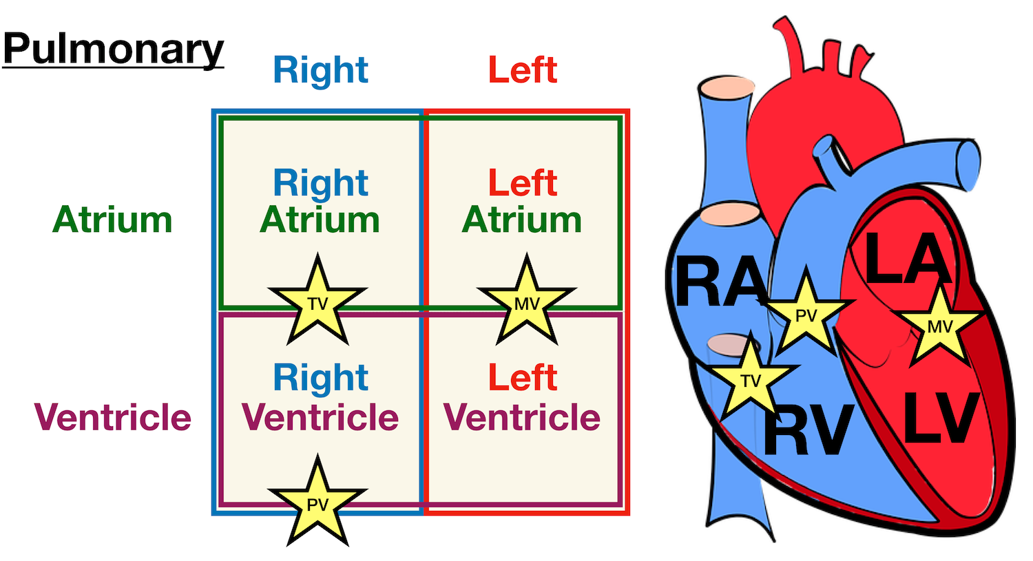
(b) blood vessels of the coronary system, including the coronary arteries and veins, keep the heart muscles oxygenated.
The heart pumps blood through a closed system of blood vessels. The heart beats continuously, pumping the equivalent of more than 14,000 litres of blood every day through five main types of blood vessels: The other arteries also thicken and stiffen. Start studying heart anatomy, blood vessels, and circulation, physiology of the heart & blood, respiratory system. Blood is carried through the body via blood vessels. The outer layer of the pericardium surrounds the roots of your heart's major blood vessels and is attached by. (1) vasculogenesis and (2) angiogenesis. The tunica externa, the tunica media, and the tunica intima. Additionally, other blood vessels return from these tissues with oxygen poor blood back to the heart. The main artery from the heart (aorta) becomes thicker, stiffer, and less flexible. Anterior heart by rogermcdougal 27,063 plays 16p image quiz. T ransports oxygen, carbon dioxide, and blood ce lls; Veins usually colored blue because oxygen poor, carry blood to the heart from the capillaries.
The outer layer of the pericardium surrounds the roots of your heart's major blood vessels and is attached by. The main artery from the heart (aorta) becomes thicker, stiffer, and less flexible. The walls of most blood vessels have three distinct layers: Anterior heart by rogermcdougal 27,063 plays 16p image quiz. Blood, heart, blood vessels ec.
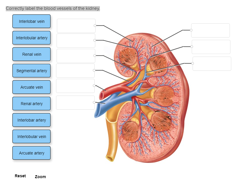
Correctly Label The Blood Vessels Of The Kidney Chegg Com from media.cheggcdn.com Start studying heart anatomy, blood vessels, and circulation, physiology of the heart & blood, respiratory system. Blood contains proteins in its red blood cells called as hemoglobin which carries oxygen to cells and tissues in the body. (1) vasculogenesis and (2) angiogenesis. It would be impossible to get blood to the predestined locations without the vascular pathways. Veins supply deoxygenated blood to the heart via inferior and superior vena cava, and it eventually drains into the right atrium. Dogfish shark heart & associated branchial blood vessels. Label the heart by lmaggieo 1,267,290 plays 21p image quiz. This is probably related to changes in the connective tissue of the blood vessel wall.
Start studying heart anatomy, blood vessels, and circulation, physiology of the heart & blood, respiratory system.
It would be impossible to get blood to the predestined locations without the vascular pathways. (a) the heart is primarily made of a thick muscle layer, called the myocardium, surrounded by membranes. The main artery from the heart (aorta) becomes thicker, stiffer, and less flexible. Structure and function of blood vessels. The other arteries also thicken and stiffen. Blood is carried through the body via blood vessels. Anterior heart by rogermcdougal 27,063 plays 16p image quiz. The following are some of the blood vessels associated with the heart: Oxygenated blood flows away from the heart through arteries. Learn vocabulary, terms, and more with flashcards, games, and other study tools. It carries oxygenated blood (pumped by the left side of the heart) to the rest of the body. The heart pumps blood through a closed system of blood vessels. Blood vessels are intricate networks of hollow tubes that transport blood throughout the entire body.
Veins usually colored blue because oxygen poor, carry blood to the heart from the capillaries. It carries oxygenated blood (pumped by the left side of the heart) to the rest of the body. Ventral views of hearts with ventral aorta & afferent arterioles: The main artery from the heart (aorta) becomes thicker, stiffer, and less flexible. Blood vessels form the extensive networks by which blood leaves the heart to supply tissue.
Circulatory System Anatomy Coronary Circulation Arteries And Cardiac Veins Vessel Model Description Youtube from i.ytimg.com The main artery from the heart (aorta) becomes thicker, stiffer, and less flexible. Ventral view of heart with ventricle pulled anteriorly to expose part of sinus venosus. Blood, heart, blood vessels ec. Your heart is located between your lungs in the middle of your chest, behind and slightly to the left of your breastbone (sternum). Arteries usually colored red because oxygen rich, carry blood away from the heart to capillaries within the tissues. The largest artery in the body, of which most major arteries branch off from. It would be impossible to get blood to the predestined locations without the vascular pathways. Veins usually colored blue because oxygen poor, carry blood to the heart from the capillaries.
Labeled shark heart & ventral aorta. Blood is carried through the body via blood vessels. Ventral view of heart with ventricle pulled anteriorly to expose part of sinus venosus. Oxygenated blood flows away from the heart through arteries. The cardiovascular system consists of the heart, blood vessels, and the approximately 5 liters of blood that the blood vessels transport. Blood vessel formation occurs via two main mechanisms: The major (or great) blood vessels of the heart are the larger arteres and veins that attach to the atria and ventricles and transport blood to and from the systemic circulatory system and the pulmonary circulatory system. Additionally, other blood vessels return from these tissues with oxygen poor blood back to the heart. Blood vessels form the extensive networks by which blood leaves the heart to supply tissue. The heart is a muscular pump that pushes blood through blood vessels around the body. They are the main blood vessels that carry the deoxygenated venous blood from the rest of the body to the right side of the heart, specifically the right atrium. (a) the heart is primarily made of a thick muscle layer, called the myocardium, surrounded by membranes. The superior vena cava is located superiorly, and it carries the deoxygenated venous blood from the upper body to the right atrium.
The tunica externa, the tunica media, and the tunica intima blood vessels labeled. Anatomy of the heart labeled diagram showing the main cardiac structures including the aorta.